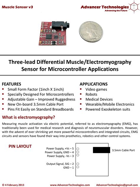
What are EMG Sensors?
EMG sensors are devices that detect and record the electrical signals generated by muscles when they contract or relax. These signals, known as myoelectric signals, are the result of the coordinated activity of motor units within the muscle. A motor unit consists of a motor neuron and the muscle fibers it innervates. When a motor neuron fires, it causes the muscle fibers to contract, generating an electrical potential that can be detected by EMG sensors placed on the skin’s surface.
Types of EMG Sensors
There are two main types of EMG sensors:
1. Surface EMG (sEMG) Sensors
Surface EMG sensors are non-invasive and are placed on the skin’s surface above the muscle of interest. They consist of electrodes that detect the electrical signals generated by the underlying muscle. sEMG sensors are widely used in research, sports science, and rehabilitation due to their ease of use and non-invasive nature.
2. Intramuscular EMG (iEMG) Sensors
Intramuscular EMG sensors are invasive and involve inserting a needle or fine-wire electrode directly into the muscle. These sensors can provide more precise and localized measurements of muscle activity compared to sEMG sensors. However, due to their invasive nature, iEMG sensors are primarily used in clinical settings and research involving deep or small muscles.
How Do EMG Sensors Work?
The working principle of EMG sensors involves detecting the electrical signals generated by muscles and converting them into a measurable output. Here’s a step-by-step breakdown of how EMG sensors work:
1. Muscle Activation
When a muscle contracts or relaxes, the motor units within the muscle generate electrical signals called action potentials. These action potentials propagate along the muscle fibers, causing them to contract.
2. Signal Detection
EMG sensors, either surface or intramuscular, detect the electrical signals generated by the muscle. The sensors are placed on the skin’s surface or inserted into the muscle, depending on the type of sensor used.
3. Signal Amplification
The electrical signals detected by the EMG sensors are typically very small, ranging from microvolts to millivolts. To make these signals measurable, they need to be amplified. EMG sensors use differential amplifiers to amplify the signal while rejecting common-mode noise, such as electromagnetic interference from nearby electrical devices.
4. Signal Filtering
After amplification, the EMG signal is filtered to remove unwanted noise and artifacts. Various filters, such as high-pass, low-pass, and notch filters, are used to remove baseline drift, movement artifacts, and power line interference.
5. Analog-to-Digital Conversion
The filtered analog EMG signal is then converted into a digital signal using an analog-to-digital converter (ADC). This digital signal can be easily processed, stored, and analyzed using computer software.
6. Signal Processing and Analysis
The digital EMG signal undergoes further processing and analysis to extract meaningful information. Common signal processing techniques include rectification, smoothing, and normalization. These techniques help to quantify muscle activity, identify patterns, and compare signals across different muscles or individuals.

Applications of EMG Sensors
EMG sensors have a wide range of applications in various fields, including:
1. Medical Diagnostics
EMG sensors are used in medical diagnostics to assess the health and function of muscles and nerves. They can help diagnose neuromuscular disorders, such as muscular dystrophy, amyotrophic lateral sclerosis (ALS), and carpal tunnel syndrome. EMG tests involve measuring the electrical activity of muscles at rest and during contraction, as well as the nerve conduction velocity.
2. Rehabilitation and Physical Therapy
EMG sensors are used in rehabilitation and physical therapy to monitor muscle activity and provide biofeedback to patients. By visualizing the EMG signals, patients can learn to control and strengthen specific muscles, which is particularly useful in treating conditions such as stroke, spinal cord injuries, and musculoskeletal disorders.
3. Prosthetic Control
EMG sensors are used in myoelectric prostheses to control artificial limbs. By detecting the EMG signals from the remaining muscles in the residual limb, prosthetic devices can be controlled in a more intuitive and natural way. This allows amputees to perform tasks such as grasping objects, opening and closing the hand, and even individual finger movements.
4. Human-Computer Interaction
EMG sensors are used in human-computer interaction (HCI) to create novel input devices and control systems. By measuring the EMG signals from specific muscles, users can control computers, video games, and other electronic devices hands-free. This has applications in accessibility, virtual reality, and augmented reality systems.
5. Ergonomics and Occupational Health
EMG sensors are used in ergonomics and occupational health to assess the physical demands of various tasks and identify potential risk factors for musculoskeletal disorders. By measuring muscle activity during work tasks, ergonomists can recommend modifications to workstations, tools, and techniques to reduce the risk of injury and improve worker comfort and productivity.
EMG Sensor Placement
The placement of EMG sensors is crucial for obtaining accurate and reliable measurements of muscle activity. The following factors should be considered when placing EMG sensors:
1. Muscle Anatomy
EMG sensors should be placed on the muscle belly, away from the tendon and motor points. The muscle belly is the thickest part of the muscle and provides the strongest EMG signal. Placing sensors too close to the tendon or motor points can result in weaker signals and increased crosstalk from neighboring muscles.
2. Electrode Orientation
The orientation of the EMG sensors should be aligned with the muscle fibers. For most muscles, this means placing the sensors along the longitudinal axis of the muscle. Incorrect orientation can lead to reduced signal amplitude and increased crosstalk.
3. Interelectrode Distance
The distance between the electrodes of an EMG sensor, known as the interelectrode distance (IED), affects the selectivity and amplitude of the recorded signal. A smaller IED provides more localized measurements but may be more susceptible to crosstalk. A larger IED provides a more global measurement of muscle activity but may be less sensitive to small changes in muscle activation.
4. Skin Preparation
Proper skin preparation is essential for obtaining high-quality EMG signals. The skin should be cleaned with alcohol to remove dirt, oil, and dead skin cells. In some cases, hair may need to be shaved to ensure good contact between the electrodes and the skin. Dry or abraded skin can lead to increased impedance and noise in the EMG signal.
EMG Signal Characteristics
The characteristics of the EMG signal provide valuable information about muscle function and health. The following are some key characteristics of the EMG signal:
1. Amplitude
The amplitude of the EMG signal represents the level of muscle activation. Higher amplitudes indicate greater muscle force or effort. The amplitude of the EMG signal can be affected by factors such as muscle size, electrode placement, and skin conductivity.
2. Frequency
The frequency content of the EMG signal reflects the firing rates of the motor units within the muscle. Healthy muscles typically have a frequency range of 20-500 Hz, with most of the signal energy concentrated between 50-150 Hz. Changes in the frequency content of the EMG signal can indicate muscle fatigue, nerve damage, or other neuromuscular disorders.
3. Timing
The timing of the EMG signal provides information about the onset, duration, and coordination of muscle activity. By comparing the timing of EMG signals from different muscles, researchers can study movement patterns, muscle synergies, and abnormalities in muscle activation.
Limitations and Challenges of EMG Sensors
While EMG sensors are valuable tools for measuring muscle activity, they have some limitations and challenges:
1. Crosstalk
Crosstalk occurs when the EMG sensor picks up signals from nearby muscles, leading to inaccurate measurements of the target muscle’s activity. Crosstalk can be minimized by careful sensor placement and using sensors with high spatial selectivity.
2. Movement Artifacts
Movement artifacts can occur when the EMG sensors or cables move relative to the skin, causing changes in the signal that are not related to muscle activity. These artifacts can be reduced by securing the sensors and cables properly and using telemetry systems to eliminate cable movement.
3. Skin Impedance
Skin impedance refers to the resistance of the skin to the flow of electrical current. High skin impedance can lead to reduced signal quality and increased noise. Proper skin preparation and the use of conductive gels can help to reduce skin impedance.
4. Signal Variability
The EMG signal can vary significantly between individuals and even within the same individual over time. Factors such as muscle size, subcutaneous fat, and skin conductivity can affect the amplitude and shape of the EMG signal. Normalization techniques, such as expressing the EMG signal as a percentage of the maximum voluntary contraction (MVC), can help to reduce variability and allow for comparisons between individuals and muscles.
Future Developments in EMG Sensor Technology
EMG sensor technology is continually evolving, with new developments aimed at improving signal quality, user comfort, and applications. Some of the future developments in EMG sensor technology include:
1. Wireless and Wearable Sensors
Wireless and wearable EMG sensors are becoming increasingly popular, as they offer greater flexibility and comfort for users. These sensors use Bluetooth or other wireless technologies to transmit the EMG signal to a receiver, eliminating the need for cumbersome cables. Wearable EMG sensors can be integrated into clothing, such as compression garments, making them suitable for long-term monitoring and real-world applications.
2. High-Density EMG Arrays
High-density EMG arrays consist of a large number of closely spaced electrodes that provide a more detailed spatial map of muscle activity. These arrays can capture the distribution of muscle activation across the entire muscle, allowing for more precise measurements and better understanding of muscle function. High-density EMG arrays are particularly useful in research settings, where detailed information about muscle activation patterns is required.
3. Machine Learning and Pattern Recognition
Machine learning and pattern recognition techniques are being increasingly applied to EMG signal analysis. These techniques can automatically detect and classify patterns in the EMG signal, such as the onset and duration of muscle activation, fatigue, and abnormalities. Machine learning algorithms can also be used to control prosthetic devices and other assistive technologies more intuitively and efficiently.
FAQ
1. Are EMG sensors safe to use?
Yes, EMG sensors are generally safe to use. Surface EMG sensors are non-invasive and do not cause any pain or discomfort. Intramuscular EMG sensors, while invasive, are used under medical supervision and with proper sterilization techniques to minimize the risk of infection.
2. Can EMG sensors be used for long-term monitoring?
Yes, EMG sensors can be used for long-term monitoring, especially with the development of wireless and wearable sensors. These sensors can be worn for extended periods, allowing for continuous monitoring of muscle activity during daily activities and sleep.
3. How do I choose the right EMG sensor for my application?
The choice of EMG sensor depends on your specific application and requirements. Factors to consider include the type of muscle being studied, the level of precision required, the need for wireless or wearable sensors, and the budget. Consulting with experts in the field and reviewing relevant literature can help guide your decision.
4. Can EMG sensors be used to control devices other than prostheses?
Yes, EMG sensors can be used to control a wide range of devices beyond prostheses. For example, EMG sensors can be used to control computer cursors, video game characters, and even drones. The field of human-computer interaction is actively exploring novel applications of EMG sensors for intuitive and hands-free device control.
5. How do I interpret EMG signal data?
Interpreting EMG signal data requires knowledge of muscle anatomy, signal processing techniques, and the specific application context. Common methods for interpreting EMG data include visual inspection of the raw signal, calculation of root mean square (RMS) or mean absolute value (MAV) amplitude, and frequency analysis using techniques such as power spectral density (PSD) and median frequency. Consultation with experts and reference to established guidelines can help ensure accurate interpretation of EMG signal data.
In conclusion, EMG sensors are powerful tools for measuring and understanding muscle activity. By detecting the electrical signals generated by muscles, EMG sensors enable a wide range of applications, from medical diagnostics and rehabilitation to prosthetic control and human-computer interaction. As EMG sensor technology continues to evolve, we can expect to see even more innovative applications and improvements in signal quality and user experience.

No responses yet